CBIS LM Core: FLUOVIEW FV3000
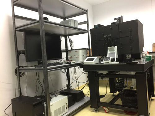
FLUOVIEW FV3000

Olympus

One new Olympus FLUOVIEW FV3000 confocal has been setup in confocal core since March 2017. FV3000 is designed not only for high sensitivity and speed imaging for live cell on 2D-6D (x,y,λ,z,t,p), but also microplate imaging and complex screening. 

LocationCBIS Light Microscopy Core
Booked CBIS equipment before? Information for First Time Users

About the FV3000

Our FV3000 is equipped with both galvanometer and high-speed resonance scanners. The resonance scanner acquires full field of view at 30 frames per second with high sensitivity and low phototoxicity, which is critical for velocity measurements and capturing dynamic events, such as for beating heart, blood flow, calcium signaling, etc. With Olympus’ exclusive silicone objectives providing clear and bright images at depth, and Olympus IX2-ZDC2, Z drift compensator, our FV3000 is optimized for time-lapse 3D imaging for culture cells.

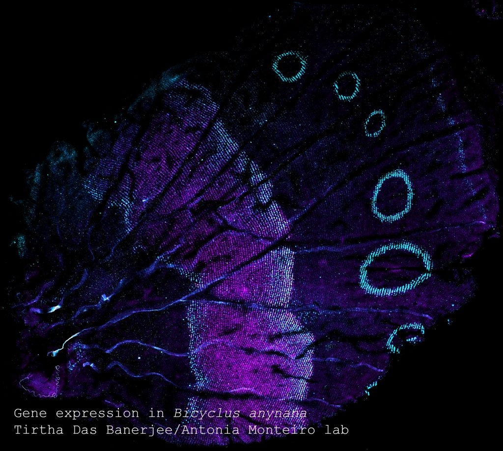
For small organisms or large pieces tissue, FV3000 facilitates macro to micro imaging at very low to high magnification (1.25X up to 100X) so that our end user is able to see the entire organism in context and focus on the interested region with high resolution information.

With Olympus’ patented Super Resolution (FV-OSR) imaging, our FV3000 provides an easy-to-use way for boosting resolution beyond the diffraction limit in fixed tissues.

Overall, the FV3000 has a range of standard and optional advanced application features including FV-OSR, spectral unmixing, photo-conversion, photo-stimulation and complex cell cycle imaging, to meet our users’ imaging requirement to answer different research questions.

Help

Features
  • Olympus IX 83 inverted microscope, with motorized XY stage IX3-SSU.
  • Environmental chamber with laser safety interlock, temperature (32C to 42C).
  • Oko lab stage insert chamber with adaptors, with CO2, humidity and temperature control
  • IX3-HO35D, 35mm dish holder and IXs-HOS, slide glass/slide chamber holder
  • Wide field light Source: coolLED pE 300 excitation system (365nm, 470nm, 565nm)
  • Filter cube on microscope  :
    • U-FUNA: Ex BP360-370, DM410, Em LP420
    • U-FBNA: Ex BP470-495, DM505, Em BP510-550
    • U-FGWA: Ex BP 530-550, DM570, Em BP575-625
  • Confocal DM:
    • DM405/488/561/640
    • DM405/488
    • DM405-445/514
    • BS10/90
    • DM 405/445/514/561/640
Objectives
  • PLANON 1.25x: Plan Apochromat Objective 1.25x/0.04, WD 5.0, Air
  • Olympus UPLSAPO10x2: U Plan Super Apochromat objective 10x/0.4, WD3.11, Air
  • Olympus UPLSAPO20x: U Plan Super Apochromat objective, 20x/0.75, WD 0.6 (spring) Air
  • UPLSAPO30xS: U Plan Super Apochromat Silicone immersion objective, 30x/1.05, WD 0.8, Silicone oil
  • Olympus UPLSAPO60xW: U Plan Apochromat Water Immersion Objective, 60x/1.2 (CC0.15-0.2), WD 0.28 Water
  • Olympus UPLSAPO60xO: U Plan Super Apochromat Objective 60x/1.35, WD 0.15 Oil
  • Olympus PLAPON60xOSC2: Super chromatic aberration correction, N.A. 1.4, WD 0.12 (chromatic aberration compensation range 405-650nm) (stand-by) Oil
Laser
  • Laser Diode : 405nm (50mW); 488nm (20mW); 561nm (20mW); 640nm (40mW)
  • Laser Diode: 445nm (75mW); 514nm (40mW)
Scanner
  • FV31-HSU-P: Hybrid scanning unit including xy galvanometer scanner set and X resonant Y galvanometer scanner. Highest scanning speed: 30fps@512×512 and 438 fps@512×32
Detector
  • FV31-SD: 2 CH spectral detector unit by Multi-Alkali PMT
  • FV31-HSD: 2CH High Sensitivity-Spectral detector unit by Cooled GaAsP PMT
  • FV31-LETD: Transmitted detector for BF/DIC
Software

    Applications
    • 2D-6D Live cell imaging: multi-channel, 3D, multiple location and time lapse imaging
    • Fixed slide imaging, tile scan
    • Advanced F-techniques: FRAP, FRET
    • Spectral Unmixing