Importance of growth factor diffusion in sensory organ development
A multi-disciplinary team consisting of NUS biophysicists along with scientists from New York University and National Institutes of Health have discovered that the protein Anosmin1 controls the diffusion of fibroblast growth factor 10 (FGF10) in the formation of sensory organs in zebrafish models.
Kallmann syndrome is a genetic disorder in humans. Patients with this condition fail to undergo puberty and have an impaired sense of smell (clinically referred to as anosmia). Mutations in the Anos1 gene (which produces the Anosmin1 protein) can cause this condition. Studies have shown that mutations in the Anos1 gene can lead to an impairment of inter-cellular communication associated with a protein called FGF10, which is a member of a family of secreted signalling proteins collectively known as fibroblast growth factors (FGFs). The FGF family, consisting of 27 different members are crucial for normal development in vertebrates. Frequently, FGFs diffuse to a different part of the tissue after they are produced to initiate various molecular development programs. However, the mechanism for regulating the diffusion of FGF10 by the Anosmin1 protein is not clear.
A research team comprising Prof Holger KNAUT and Prof Eli ROTHENBERG from New York University, Prof Martin MEIER-SCHELLERSHEIM from National Institutes of Health and Prof Thorsten WOHLAND from the Departments of Biological Sciences and Chemistry, NUS have discovered that Anosmin1 regulates and enhances the FGF10 signalling mechanism during sensory organ formation in zebrafish models by facilitating its diffusion within developing tissues. From their findings, a potential hypothesis is that mutations in the Anos1 gene can cause a reduction in the signalling range of FGF10. This in turn leads to abnormal development causing Kallmann syndrome.
The team used fluorescence correlation spectroscopy (FCS), an analytical technique to measure the diffusion and interactions of fluorescence-tagged molecules in zebrafish embryos. FCS is able to quantify the diffusion coefficient of molecules by monitoring the fluctuations in fluorescence emitted by the molecules. Their FCS studies on the FGF10 molecules showed the presence of two different types of FGF10. One of them diffuses slowly while the other diffuses at a much faster rate. The slow-diffusing type is bound to the extracellular matrix (ECM) while the fast-diffusing type diffuses freely in the zebrafish embryo. Using a related technique, fluorescence cross-correlation spectroscopy (FCCS), the team showed that Anosmin1 diffused together with FGF10. This suggests that Anosmin1 liberates FGF10 from the ECM, thus increasing the number of freely diffusing FGF10. Through this mechanism, Anosmin1 increases the diffusivity and the signalling range of FGF10 for normal development of sensory organs in zebrafish.
Prof Wohland said, “These findings are made possible with the strong collaboration between developmental biologists and biophysicists. It nicely demonstrates how single molecule sensitive techniques can provide new insights into molecular mechanisms in living organisms.”
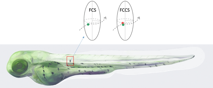
Figure shows an artistic impression of a fluorescent labelled zebrafish embryo. FCS and FCCS measurements can be conducted in any part of the fish by placing a laser focus at the corresponding point. Singly labelled molecules (green) were studied in FCS to obtain information about their diffusion and concentration. Double labelled molecules (red and green) were used in FCCS to obtain information about the interactions between them. [Credit: Sonia MONTI]
Reference
Wang J; Yin YD; Lau S; Sankaran J; Rothenberg E; Wohland T; Meier-Schellersheim M; Knaut H*, “Anosmin1 shuttles Fgf to facilitate its diffusion, increase its local concentration, and induce sensory organs” DEVELOPMENTAL CELL Volume: 46 Issue: 6 Pages: 751 DOI: 10.1016/j.devcel.2018.07.015 Published: 2018.