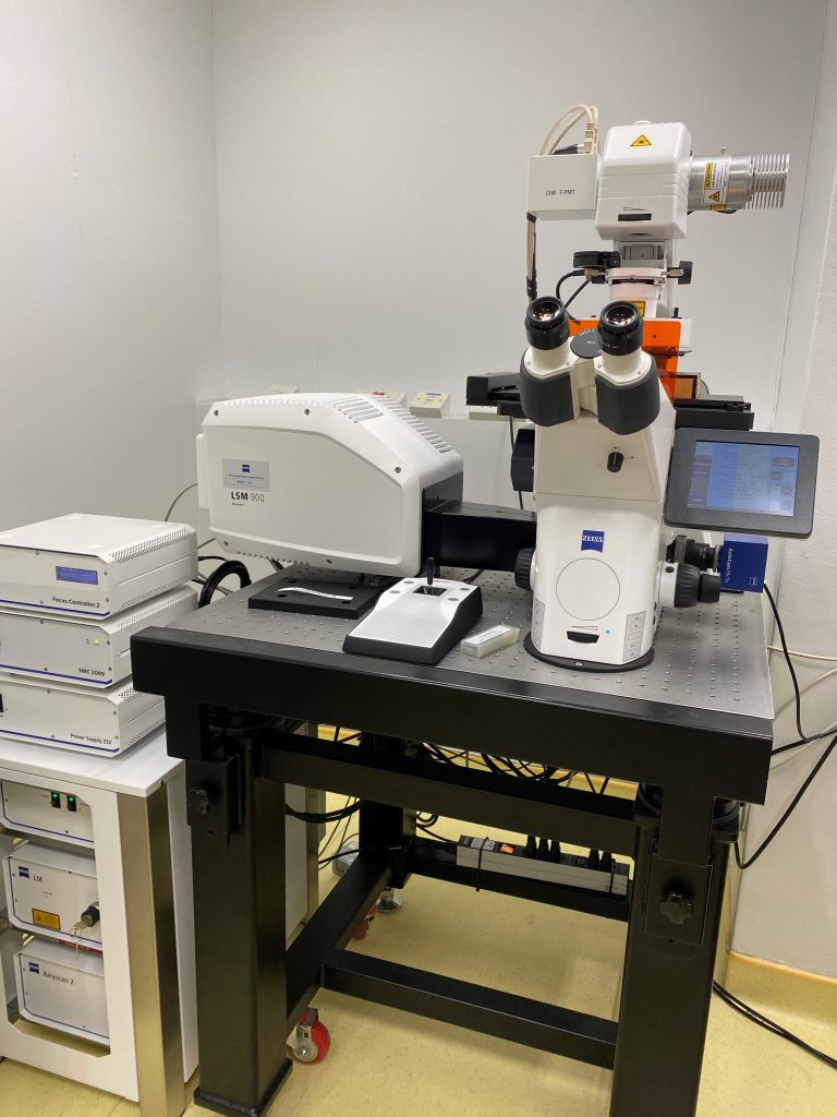
CBIS LM Core: Zeiss LSM 900 with Dynamics Profiler

LSM 900 confocal with Airyscan II and Dynamics Profiler
zeiss
The LSM 900 offers super-resolution and improved SNR via the Airyscan detector, while the Dynamics Profiler makes it easy to measure the concentration and spatial diffusion patterns of fluorescent molecules. Airyscan is a hexogen array of 32 concentrically arranged GaAsP detector; each detector approx. 0.2AU in diameter thus total detection area approx. 1.25AU to acquire most of the Airy disk all at once. The confocal pinhole itself remains open and does not block light, thus more photons are collected for the improved resolution and S/N. After detector-wise deconvolution and pixel reassignment, Airyscan can achieve 1.7x improvement over the set by the Abbe limit.
Location: S1A #03-07
Booked CBIS equipment before? Information for First Time Users
About the LSM900 Confocal with Airyscan II
Airyscan 2 has different acquisition modes: SR and SR Multiplex. For each illumination position, Airyscan SR mode generates one superresolution image pixel. The spatial information provided in the Multiplex modes SR-2Y / CO-2Y and SR-4Y allows to scan 2 or even 4 superresolution image lines in a single sweep. This allows to take bigger steps when sweeping the excitation laser over the field of view to achieve high acquisition speed with superresolution.

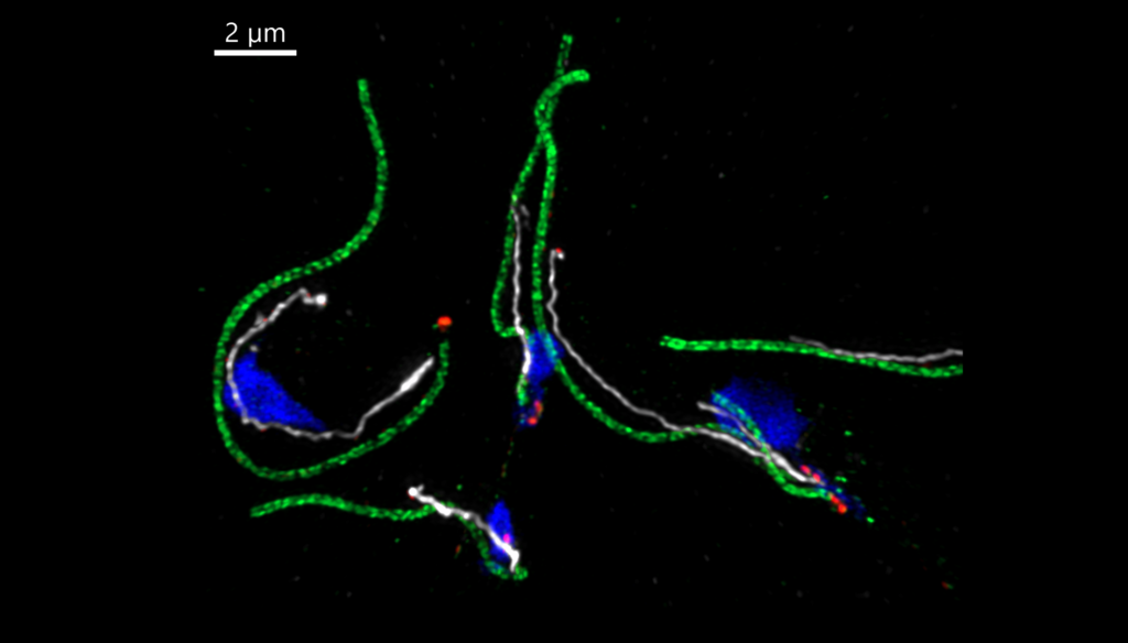
Microtubule-associated structures on the parasite Trypanosoma brucei. Green, flagellar outer arms; red, flagellum attachment complex; white, microtubule-associated protein; blue, DNA and mitochondria. Sample preparation, Assoc Prof Cynthia He; imaging, Tong Yan; Airyscan super-resolution.
With the airyscan detector, Zeiss fast linear scan and short light path, LSM900 is one of the most sensitive confocal microscopies in our core. Equipped with Zeiss Definite Focus 2, Zenblue Tile and Experiment Designer modules, LSM900 airy scan II is recommended for large area high resolution imaging.

Help
- Video: Multiplex Mode of ZEISS LSM 9 Family – AnimationMicroscope Zeiss
- Manual (Brief)
- Zen Blue Manual
Features
- Microscope: Zeiss Axio Observer 7, with motorized XY stage
- Sample holder: universal sample holder and multiple well plate holder
- Wide field fluorescent microscopy:
- Light Source: Solid-State Light Source Colibri 7, Type RGB-UV. Equipped with 4 solid state LED lamps. 630nm, 555nm, 475nm and 385nm
- Filter cube: Filter set 90 HE LED (E) for fluorescent dyes like DAPI, FITC, TRITC and Cy5 with excitation wavelengths 385, 475, 555 and 630 nm. Beam splitter QBS 405 + 493 + 575 + 653 and emission filter QBP 425/30 + 514/30 + 592/30 + 709/100.
- Scanner: Two galvanometric scanning mirrors with line and frame flyback
- Scanning speed: At 512 × 512 pixels: confocal – up to 8 fps; Airyscan SR – up to 4 fps; Multiplex SR-2Y – 8.4 fps; Multiplex SR-4Y – 18.9 fps.
- Scanning Field: 18 mm diagonal in the intermediate image plane, with full pupil illumination.
- Pinhole: Master pinhole with preset size and position; can be adjusted as desired for multitracking.
- Beampath: one Main beam splitter (MBS) for 4 laser lines (405, 488, 561 and 640nm). Two patented Variable Secondary Dichroic (VSDs) at 10 degree angle to incident beam for most effective excitation light suppression.
- XY stage: Motorized XY scanning stage, for Mark & Find function (xy) as well as Tile Scan (Mosaic Scan)
- Definite Focus.2: to stabilize the focal position.
Objectives
- Objective Plan-Apochromat 10x/0.45 M27, with DIC
- Objective Plan-Apochromat 20x/0.8 M27, with DIC
- Objective 20x/0.4 Corr LD
- Objective Plan-Apochromat 63x/1.4 Oil for SR, with DIC
- Objective Plan-Apochromat 63x/1.2 H2O for SR, with DIC
Lasers
- Laser Diode : 405nm (5mW); 488nm (10mW); 561nm (10mW); 640nm (5mW)
Detectors
- Two GaAsP (typical QE 45%) and one Airyscan detector for 63x
- Transmitted light detector
- ZEISS Axiocams: to acquire overview images or to perform light efficient widefield imaging
Software
- Zenblue 3.1 with Tile and Experiment Designer. Use the following to download Zenblue lite https://www.zeiss.com/microscopy/int/products/microscope-software/zen-lite.html
Applications
- 2D-6D Live cell imaging: multi-channel, 3D, multiple location and time lapse imaging, with airyscan super resolution using 63x lens
- Fixed slide imaging, tile scan
- FCS (fluorescence correlation spectroscopy)

