A History of the NUS Centre for Bioimaging Sciences
An interdisciplinary research centre for leading-edge microscopy & computatioN
Founded in 2008, and opened in its current premises in 2010, CBIS was created as an interdisciplinary research centre combining existing groups in light, electron, and scanning probe microscopy into a centre to study the basic structure and dynamics of cellular systems over wide temporal and spatial ranges. CBIS acts especially in areas that offer collaboration with large-scale research programs such as Cancer RCE, SBIC, SMART Infectious Disease IRG, and mechanobiology.
Original whitepaper for the founding of CBIS (PDF)
 About the Centre
CBIS’s Objective: to bring together an interdisciplinary group of biologists, chemists, computer scientists, mathematicians and physicists, all with a common belief; that complex cellular processes can be probed and captured with sophisticated imagining techniques, and then understood through powerful computational methods.
CBIS’s Mission by Founding Director, Paul Matsudaira
Through the centuries, discoveries have built on discoveries, and knowledge has built on knowledge. Technology has opened avenues of exploration never before conceived. Then there is the present. We are living in an exciting time, when we have both the awareness and expertise to explore ever-expanding scales of knowledge about molecules and cells, and the technology to make it possible. In particular, advances in imaging have catalyzed advances in biology. The NUS Centre for BioImaging Sciences was founded to develoop and apply novel imaging technologies to key problems in biology.
Recent advances in biology, chemistry, computer sciences, and physics –and the remarkable evolution of microscopes and computation– enable us to watch single molecules in action, create three-dimensional images of molecules over time, and observe them in live cells and tissues. Biomolecular imaging has not only gone far beyond its earlier capabilities, it has also surpassed other traditional methods of cellular research. Now we have the means to delve into systems-scale questions, observing many different molecules – and the structures they comprise – simultaneously and in real time.
CBIS is working to find new answers as well as ask new questions that enhance our understanding of the most complex issues in biology. We invite you to explore the Centre for BioImaging Sciences, and to join us in applying our tools, resources, and expertise to the microscopic mysteries that await discovery.
History of CBIS’ CryoEM Facility
The department first introduced electron microscopy at NUS in 1984 when the Government of Japan presented a JEOL JEM 100 CXII STEM equipped with X-ray Microanalysis attachment and other sample preparation equipment. It was at that time the most up-to-date facility in Singapore and it served the whole of the Science Faculty and other institutes, such as SGH.
This equipment analysed a wide range of biological and physical samples from unicellular organisms to tissues of both botanical and zoological nature and from nanoparticles to geological samples. It continues to be used by research personnel from many faculties and departments of NUS and industry.
Challenges to building the new facility
Prior to renovation, the Microscopy Facility experienced two major problems. The high relative humidity of the tropics caused instantaneous ice crystal formation on cryo-samples. This spurious ice contaminant coated the cryosamples and prevented structure determination in the electron microscope.
An equally serious problem was that a nearby building power and ground cables generated extremely high electromagnetic interference, EMI, which distorted the electron beam of all the TEMs and prevented them from reaching their full performance specifications.
JEOL and FEI engineers determined the sources of the EMI and contributed to the design of the Cryoelectron Microscopy Facility. As a result, the Facility was designed to support high-end cryoelectron microscopy by reducing humidity to 30% RH in the microscope and sample preparation rooms and by minimizing EMI by moving the power and ground lines for buildings S1, S2, S3, and S4 a suitable distance away from the Cryo Facility
After renovation, the Microscopy Facility was reorganized into the Cryoelectron Microscopy Facility by moving non-cryo-related functions to other sites including a scanning electron microscope to the TMSI and light microscopy support to building S1A.
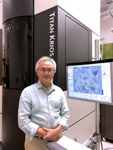
Acquisition of the Titan Krios
The flagship instrument of the Facility is the FEI Titan Krios. Fully automated, the microscope lies within an outer box to give its iconic shape and shield the microscope from fluctuations in temperature and acoustic noise.
An autoloader feeds up to twelve samples into the specimen holder for imaging at liquid nitrogen temperatures. The microscope is operated remotely from a station located outside the microscope room. Images are collected by a 4K x 4K Gatan CCD camera after post-filtering through a Gatan GIF. Shortly after installation, the Krios was collecting high-resolution images of viruses and other biological specimens. This multi-million dollar instrument was purchased by funds from the Lee Hiok Kwee Fund.
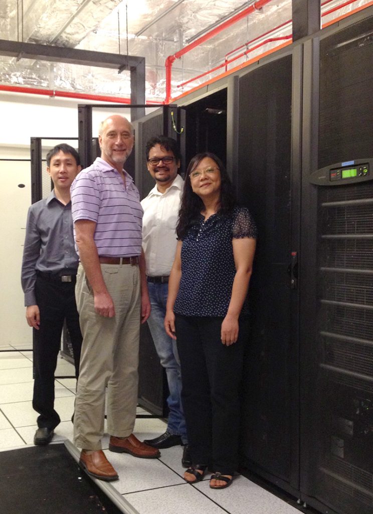 History of the CBIS Computational Facility
Core Mission by founding architect, Alan Davis: CBIS’s computational core facility was created to store, process, analyse and visualise experimental data and images, as well as large scale computer modeling of cellular processes and data mining of complex biological data.
Challenges
Since the CBIS instruments can collect very large data sets approaching 1 TB in size, all IT infrastructure was designed to accommodate the movement and storage of large data sets during their acquisition, analysis and long term storage phases. Typical scientific laptops and workstations used by researchers, along with the local networks and storage systems, are not capable of handling this amount of data. In addition, the university general purpose computational facilities were unable to be used without invoking large time delays in data movement, restrictions in data set sizes, and offline batch computing analysis unsuitable for modern scientific research.
Facility Design
As CBIS moved to a new research space without any preexisting IT facilities, a new state of the art data centre was designed to accommodate the high performance computational, storage and networking capabilities planned for the facility. These IT resources were designed as a follow on to similar facilities designed by Alan Davis for Paul Matsudaira’s BioImaging Center and the Computational Systems Biology Institute at MIT in Cambridge, MA, although they were extensively updated after consultations with the CBIS researchers to accommodate their specific requirements and new instrumentation. CBIS researchers had designed a new Lightsheet Microscope capable of collecting data at very high data rates compared with traditional OMs, and would house multiple EMs compared to what was in use at MIT. This enhanced data collection capability meant all aspects of the IT resources would need updating for these increased data collection rates, storage capacity and data processing requirements.
IT Resources
The initial general purpose networking resource included 10 Gigabit Ethernet (GbE) connections not only within the data center but also to all microscope systems and individual workstations. This network has subsequently been upgraded to 40 GbE within the data center and to the EM facility.
The storage system initially consisted of a 100+ TB high performance storage array using Hierarchical Storage Management software to automatically manage the data. A dedicated high speed network was installed between the main storage array and the HPC servers to offload this data traffic from the slower general purpose network. This system was later expanded to 500 TBs of capacity with an archival tier. After many years in use, a new 1.2 BP storage array was installed utilizing InfiniBand (IB) networks as a replacement. This system is now in the process of being replaced by a 2 PB storage array to accommodate the ever expanding data storage requirements. Thus the storage capacity has increased 20-fold with a 5-fold increase in performance over the short lifetime of CBIS.
The compute servers initially included two multi-core SGI shared memory servers with > 100 TB of shared memory to facilitate applications that could utilize both many cores and large amounts of memory. Subsequently, this was augmented with two multi-node clusters, one with GPUs for applications that could use them for accelerated computational speed. The clusters are interconnected with a high speed IB network to facilitate applications that can run on multiple nodes. This has resulted in a 100-fold increase in computational performance since the start of CBIS.