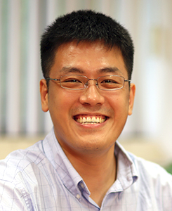
LU GAN
Associate Professor
Contact Information:
Department of Biological Sciences
National University of Singapore
14 Science Drive 4
Singapore 117543
6516 8868
6779 2486
lu@anaphase.org
Academic Qualifications
Postdoc, California Institute of Technology, 2011
Ph.D., The Scripps Research Institute, 2006
B.S., California Institute of Technology, 2001
Research Interests
Chromosomes are the first and final frontier of structural cell biology. They contain the instructions of life, which must be read, copied, and passed on to daughter cells. These activities must be done accurately by each of the trillions of cells in the human body. Mistakes can lead to many diseases, including cancers. How do cells access and use the information stored in chromosomes? How do they fold up the fundamental subunits called nucleosomes and then transport them into daughter cells? The answers to many of these fundamental questions will require knowledge of the organization, positions, and distributions of the subunits of chromosomes and their interaction partners, all within their native intracellular context.
The DNA and proteins that make up chromosomes are measured in nanometers, but they reside inside cells that are measured in micrometers. To map out the nanoscale organization of chromosomes inside their native cellular context, we use state-of-the-art electron tomography to generate 3-D images called “tomograms”. To minimize the fixation, dehydration, extraction, and staining artifacts associated with conventional electron microscopy (EM), we prepare and image our cells in a life-like frozen-hydrated state. This combination of 3-D nanoscale imaging and cryogenic sample handling is called cryo-electron tomography (cryo-ET). All our work has been done at the NUS Center for Bioimaging Sciences, which is equipped with top-of-the-line instruments, such as a 300-keV electron cryomicroscope (cryo-EM), the Titan Krios. Images are acquired on a direct-detection camera, the Falcon II, which has contributed to the cryo-EM revolution that is currently underway.
To answer questions about chromosome biology using cryo-ET, we have chosen budding and fission yeasts as our model organisms. Yeasts have numerous advantages, including their simplified cytology, simplified gene structure, and ease of growth and maintenance. The heroic efforts of our former PhD student Chen Chen have made cryo-ET of yeast routine. We have now exploited yeast to ask how cell-cycle, post-translational modifications, and architectural proteins influence chromosome structure.
Current Projects
Chromosome organization in unicellular eukaryotes
Chromosome organization at the nucleosome level is thought to regulate access to key control sequences and protein interfaces. The textbook 30-nm fiber model explains how this happens: sequential nucleosomes fold into a dense ~ 30-nm-diameter fiber. This close packing hides the “face” of each cylinder-shaped nucleosome and at least half the DNA wrapped around it. For the past four decades, such fibers were seen nearly universally when chromosomes were purified and then digested into small fragments. In 1986, the pioneering in situ cryo-EM work of McDowall, Smith and Dubochet led to the alternate “liquid” model in which there is no long-range order within a chromosome. Technological advances in the past decade have allowed other groups and ours to revisit this question. We have shown that 30-nm fibers cannot explain any of the chromosomes visualized in picoplankton or budding yeast, in either interphase or mitosis (Gan, 2013; Chen, 2016). The best model that can explain chromosome folding is a disordered zig-zag of nucleosomes. There is no global condensation and or long-range order of any kind. In yeast, this “open” configuration facilitates access to promoters and allows for high levels of transcription throughout the cell cycle.
What are the factors that control chromosome organization? This question may take decades to answer because there are many biophysical factors (crowding, charge, polymer stiffness) and cellular factors (histone variants, transcription, architectural proteins). Addressing each of these factors – and combinations thereof – in vivo is presently unfeasible because the time-consuming cellular sample preparation is not automated. We have therefore developed a simple method to obtain tomograms of chromosomes in vitro, but closer to the natural state: we lyse cells or nuclei and then image the nuclear contents before they can be damaged by protease and nucleases (Cai, 2017). These lysates allow us to ask how much charge contributes to chromatin organization. Picoplankton chromosomes respond to divalent cations (like magnesium) in a somewhat-predictable fashion: they unfold into open zigzags when deprived of magnesium, but clump up into large amorphous masses when treated with low concentrations of magnesium. To our surprise, yeast chromosomes are largely compacted as a large mass, even in the absence of free magnesium and when treated a drug that makes chromosomes more negatively charged. Furthermore, 30-nm fibers are much rarer in either of these samples than expected. Because undigested chromosomes constrain nucleosome concentration to be much higher than digested chromosome fragments, our findings support the “liquid / polymer-melt” models proposed by Eltsov, Dubochet, and colleagues.
Nucleosome rearrangement in chromosome condensation
Chromosome condensation is a hallmark of dividing cells, and has been known for more than a century. We have recently used cryo-ET to ask how nucleosomes are rearranged in dividing fission yeast, a simple cell whose chromosomes compact into discrete bodies just like in human cells. In textbook models, chromosomes condense by the uniform accretion of all their nucleosomes. Instead, we found that condensed chromosomes are not uniform. There are large macromolecular complexes the size of ribosomes and numerous nucleosome-free pockets, all embedded within the chromosome; both features are also present in interphase, where they are more abundant. Chromosomes are therefore porous throughout the cell cycle. Indeed, we were able to confirm that active RNA polymerases exist inside. Therefore, at least in dividing fission yeast cells, chromosomes are not metabolically inert bodies, but are active in transcription and perhaps other activities yet to be discovered (Cai, 2018).
Please visit our website for more updates, including newer preprints.
Selected Publications and Preprints
-
Ng, C., Deng, L., Chen, C., Lim, H.H., Shi, J., Surana, U., Gan, L. (2018), Electron cryotomography analysis of Dam1C/DASH at the kinetochore-spindle interface in situ. Journal of Cell Biology, 2019.
-
Cai, S., Chen, C., Tan, Z.Y., Huang, Y., Shi, J., Gan, L. (2018), Cryo-ET reveals nucleosome reorganization in condensed mitotic chromosomes in vivo. Proceedings of the National Academy of Sciences 115: 10977-10982
-
Cai, S.†, Böck, D.†, Pilhofer, M.*, Gan, L.* (2018), The in situ structures of mono-, di-, and trinucleosomes in human heterochromatin. Molecular Biology of the Cell 29: 2450-2457 († equal contributions); * corresponding authors
-
Cai, S., Song, Y., Chen, C., Shi, J., Gan, L. (2018), Natural chromatin is heterogeneous and self-associates in vitro. Molecular Biology of the Cell 29: 1652-1663
-
Chen, C., Lim, H.H., Shi, J., Tamura, S., Maeshima, K., Surana, U., Gan, L. (2016), Budding yeast chromatin is dispersed in a crowded nucleoplasm in vivo. Molecular Biology of the Cell 27: 3357-3368
