Epidermal Growth Factor Receptor
Cell signalling governs and coordinates multiple cell activities, including proliferation and development. Receptor Tyrosine Kinases (RTKs) are a family of bitopic, cell surface receptor proteins, which transduce signals through phosphorylation of intracellular tyrosine residues. The subclass I of the RTK superfamily is the ErbB (or HER) family, containing four members (ErbB1-4). The Epidermal Growth Factor Receptor (EGFR/ErbB1/HER1) is one of the best-studied receptor proteins. Nevertheless, a clear understanding of how the receptor is activated and its fate after activation has been elusive. Since EGFR mutations and dysregulated signalling have been implicated in many cancers, it is important to understand the oligomerization and dynamics of EGFR on the plasma membrane [1]. For some recent reviews, please refer [2–4].
Our previous work
The presence of preformed EGFR dimers in the absence of EGF has been reported by numerous studies [5]. Our lab has also previously studied preformed dimers of EGFR, mainly utilising fluorescence cross-correlation spectroscopy (FCCS). Liu et al. showed preformed homo- and heterodimers of EGFR and ErbB2 in resting CHO-K1 cells using single-wavelength FCCS (SW-FCCS) and Förster resonance energy transfer (FRET) [6]. Ma et al. confirmed the presence of preformed EGFR dimers and showed the interaction of phosphotyrosine binding domain (PTB) with EGFR before and after EGF stimulation using SW-FCCS [7]. Yavas et al. studied the effects of various experimental factors like cell lines with different EGFR expression levels, temperature, and membrane side on EGFR dimerization in resting cells using various FCCS modalities [5]. Bag et al. studied the lateral diffusion and organization of EGFR on the plasma membrane of CHO-K1 cells using imaging total internal reflection-fluorescence correlation spectroscopy (ITIR-FCS) [8].
Our current focus
The dimerization state of EGFR before activation remains to be understood better and higher-order oligomers have not been quantified. In addition, the components of the plasma membrane are involved in the regulation of EGFR, but these interactions have not been sufficiently studied. A more comprehensive understanding of these interactions is required. We are currently using various drug treatments and EGFR mutants to answer these questions, utilising Number and Brightness (N&B) analysis.

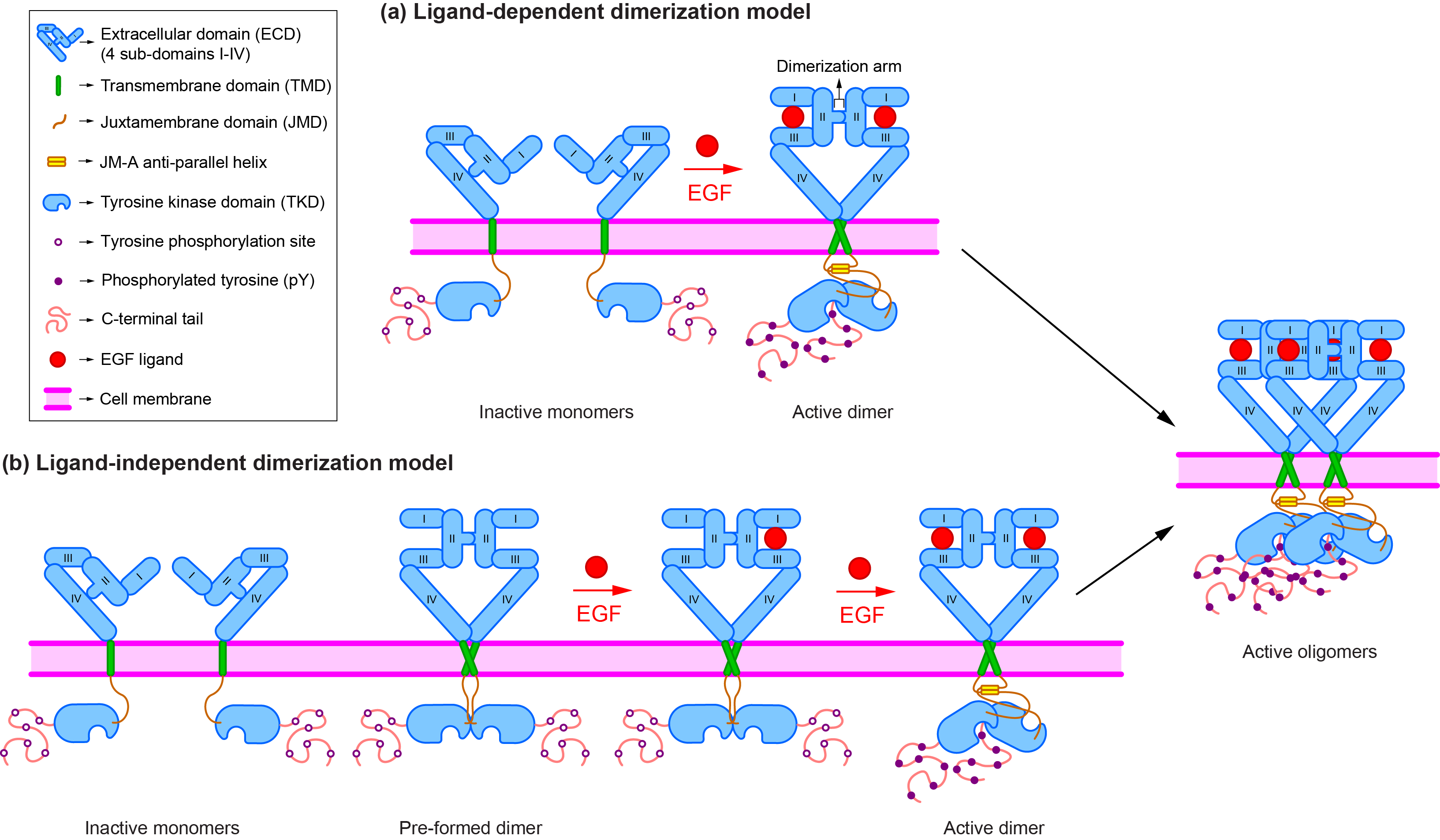
The canonical ligand-dependent dimerization model proposes that EGFR (shown in blue) exists as monomers on the cell membrane prior to ligand (shown in red) binding. It changes its conformation from the tethered to the extended form upon ligand binding and dimerizes with another EGFR molecule. On the other hand, the alternative ligand-independent model takes into consideration the presence of preformed dimers. This model proposes that the preformed dimers are inactive and adopt an untethered, symmetric structure. Upon ligand binding, the kinase domains are reoriented from the inactive, symmetric structure into the active, asymmetric structure. Moreover, it has been further argued that EGFR dimers are not sufficient and higher-order oligomers (like tetramers) are required for full receptor activation and signalling.
References
1. Maruyama I. Mechanisms of Activation of Receptor Tyrosine Kinases: Monomers or Dimers.
Cells. 2014;3(2):304–30.
2. Kovacs E, Zorn JA, Huang Y, Barros T, Kuriyan J. A structural perspective on the
regulation of the epidermal growth factor receptor. Annu Rev Biochem. 2015;84:739–64.
3. Endres NF, Barros T, Cantor AJ, Kuriyan J. Emerging concepts in the regulation of the
EGF receptor and other receptor tyrosine kinases. Trends Biochem Sci. 2014;39(10):437–46.
4. Bocharov E V., Sharonov G V., Bocharova O V., Pavlov K V. Conformational transitions
and interactions underlying the function of membrane embedded receptor protein kinases. Biochim
Biophys Acta - Biomembr. 2017;1859(9):1417–29.
5. Yavas S, Macháň R, Wohland T. The Epidermal Growth Factor Receptor Forms
Location-Dependent Complexes in Resting Cells. Biophys J. 2016;111(10):2241–54.
6. Liu P, Sudhaharan T, Koh RML, Hwang LC, Ahmed S, Maruyama IN, et al. Investigation of
the dimerization of proteins from the epidermal growth factor receptor family by single
wavelength fluorescence cross-correlation spectroscopy. Biophys J. 2007;93(2):684–98.
7. Ma X, Ahmed S, Wohland T. EGFR activation monitored by SW-FCCS in live cells Xiaoxiao.
Front Biosci (Elite Ed). 2011;E3:22–32.
8. Bag N, Huang S, Wohland T. Plasma Membrane Organization of Epidermal Growth Factor
Receptor in Resting and Ligand-Bound States. Biophys J. 2015;109(9):1925–36.

